

JPNTestからの試験合格準備の必需品SPI試験トレーニング問題
有効なパス率はARDMS SPIのSPI試験問題
ARDMS SPI 認定試験の出題範囲:
| トピック | 出題範囲 |
|---|---|
| トピック 1 |
|
| トピック 2 |
|
| トピック 3 |
|
| トピック 4 |
|
| トピック 5 |
|
質問 # 56
The calipers in this image measure which performance characteristic of a system?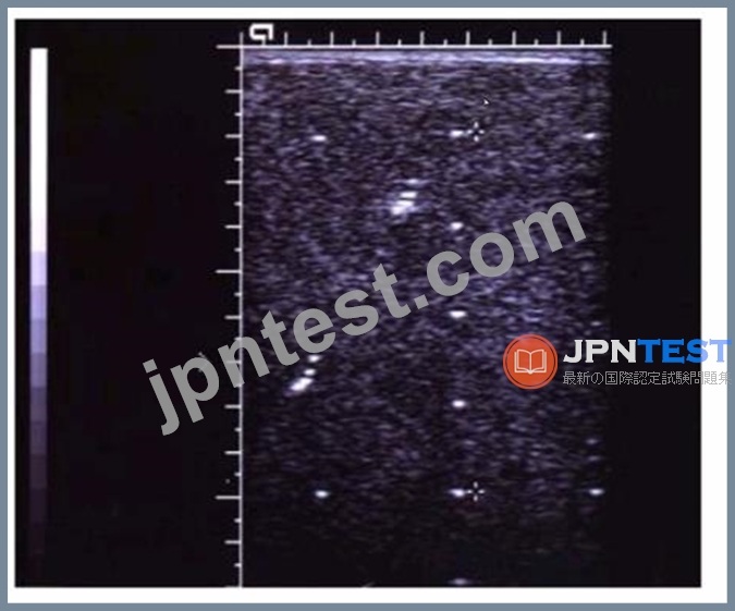
- A. Depth measurement accuracy
- B. Lateral resolution
- C. Dynamic range
- D. Axial resolution
正解:A
解説:
The calipers shown in the image are used to measure the depth of structures within the ultrasound image. This performance characteristic, known as depth measurement accuracy, assesses how accurately the ultrasound system can measure the distance from the transducer to a specific point within the body. Accurate depth measurements are crucial for diagnostic purposes, ensuring that anatomical and pathological structures are correctly identified and evaluated.
Reference:
American Registry for Diagnostic Medical Sonography (ARDMS) Sonography Principles and Instrumentation study materials.
Textbook of Diagnostic Sonography by Hagen-Ansert, S. L. (latest edition).
質問 # 57
Which target group in this image of a tissue-mimicking phantom is used to evaluate axial resolution?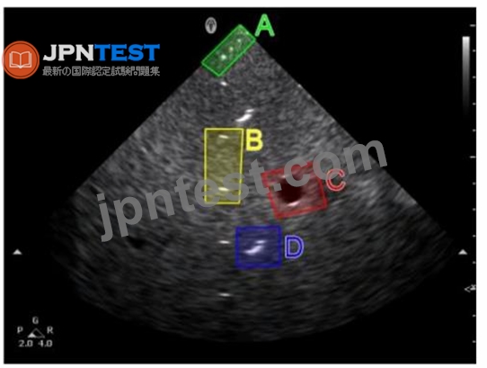
- A. Option B
- B. Option D
- C. Option C
- D. Option A
正解:A
解説:
In the given image of a tissue-mimicking phantom, Option B (yellow box) is used to evaluate axial resolution. Axial resolution refers to the ability of the ultrasound system to distinguish between two structures that are close to each other along the path of the ultrasound beam (i.e., parallel to the beam). The targets in Option B are typically aligned in such a way to test the system's capacity to differentiate between structures that are situated closely together along the beam's axis. Reference:
ARDMS Sonography Principles and Instrumentation guidelines
"Sonography: Principles and Instruments" by Joan P. Baker and Marveen Craig
質問 # 58
Which pulsed-wave Doppler adjustment would be appropriate to correct the aliasing seen in this image?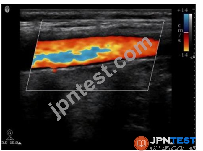
- A. Decrease the Doppler pulse repetition frequency.
- B. Decrease the spectral Doppler gain.
- C. Increase the spectral Doppler gain.
- D. Increase the Doppler pulse repetition frequency.
正解:D
解説:
Aliasing in pulsed-wave Doppler occurs when the sampled Doppler frequency exceeds the Nyquist limit, which is half of the pulse repetition frequency (PRF). This results in an incorrect representation of the blood flow velocities, causing the waveform to wrap around and appear on the opposite side of the baseline. To correct aliasing, the PRF should be increased, which raises the Nyquist limit and allows for accurate measurement of higher velocities without aliasing. Increasing the PRF effectively reduces the likelihood of aliasing artifacts in the Doppler signal.
Reference:
American Registry for Diagnostic Medical Sonography (ARDMS). Sonography Principles and Instrumentation (SPI) Examination Review Guide.
質問 # 59
What produces increased attenuation within soft tissue?
- A. Higher intensity of the ultrasound beam
- B. Lower intensity of the ultrasound beam
- C. Higher frequency of the ultrasound beam
- D. Lower frequency of the ultrasound beam
正解:C
解説:
Attenuation refers to the reduction in the intensity of the ultrasound beam as it travels through tissue. Higher frequency ultrasound beams experience more attenuation because they are absorbed and scattered more than lower frequency beams. This is due to the fact that higher frequency waves have shorter wavelengths and interact more with the small particles in tissues, causing greater energy loss.
Reference: ARDMS Sonography Principles and Instrumentation, Chapter on Ultrasound Physics and Instrumentation.
質問 # 60
Which adjustment follows the ALARA (As Low As Reasonably Achievable) principle of patient safety?
- A. Increase number of focal zones
- B. Increase sector width
- C. Decrease output power
- D. Decrease depth
正解:C
解説:
The ALARA principle stands for "As Low As Reasonably Achievable" and aims to minimize patient exposure to ultrasound energy. Decreasing the output power reduces the intensity of the ultrasound waves, thereby reducing the potential for tissue heating and mechanical effects, which aligns with the ALARA principle of minimizing exposure while still obtaining necessary diagnostic information.
Reference:
ARDMS Sonography Principles and Instrumentation guidelines
Hangiandreou, N. J. (2003). "Physics Tutorial for Residents: Topics in US: B-Mode US: Basic Concepts and New Technology." Radiographics.
質問 # 61
What is a potential negative consequence of using a high wall filter?
- A. Desired signal may be eliminated
- B. Penetration is reduced
- C. Too much noise may appear on the image
- D. Aliasing could occur
正解:A
解説:
A high wall filter is used in Doppler ultrasound to eliminate low-frequency signals that may be attributed to vessel wall motion or other low-velocity flows. However, if the wall filter is set too high, it can inadvertently eliminate desired low-frequency Doppler signals that represent real blood flow, particularly in smaller vessels or those with slower flow velocities. This results in a loss of valuable diagnostic information.
Reference: ARDMS Sonography Principles and Instrumentation (SPI) Review, Doppler Ultrasound section.
質問 # 62
Which describes the reflected frequency when a reflector is moving toward the sound source?
- A. Decreased
- B. Attenuated
- C. Unchanged
- D. Increased
正解:D
解説:
When a reflector (such as red blood cells) is moving toward the sound source, the frequency of the reflected sound waves increases. This phenomenon is known as the Doppler effect. The frequency shift occurs because the motion of the reflector compresses the sound waves, leading to a higher frequency than the emitted frequency. This increased frequency is what the Doppler ultrasound system detects and uses to calculate the velocity of the moving reflector.
Reference:
ARDMS Sonography Principles and Instrumentation guidelines
Hoskins, P. R., Thrush, A., Martin, K., & Whittingham, T. A. (2010). Diagnostic Ultrasound: Physics and Equipment.
質問 # 63
What is the term for an ultrasound system's ability to display low-level echoes?
- A. Sensitivity
- B. Lateral resolution
- C. Slice thickness
- D. Axial resolution
正解:A
解説:
Sensitivity is the term for an ultrasound system's ability to display low-level echoes. It refers to the system's capacity to detect and accurately display weak echoes returning from tissues. High sensitivity allows the sonographer to visualize structures that produce faint echoes, such as small or low-contrast lesions. This parameter is critical for ensuring that subtle pathological changes are not missed during imaging. Reference:
ARDMS Sonography Principles and Instrumentation guidelines
"Sonography: Principles and Instruments" by Joan P. Baker and Marveen Craig
質問 # 64
Which resolution is improved by focusing?
- A. Axial
- B. Contrast
- C. Lateral
- D. Temporal
正解:C
解説:
Focusing improves lateral resolution in ultrasound imaging. Lateral resolution refers to the system's ability to distinguish between two points that are side by side (perpendicular to the sound beam's path). By focusing the ultrasound beam, the width of the beam is narrowed at the focal point, enhancing the system's ability to resolve structures that are close together in the lateral plane. This results in clearer, more detailed images of the anatomical structures.
Reference:
American Registry for Diagnostic Medical Sonography (ARDMS) Sonography Principles and Instrumentation study materials.
Diagnostic Ultrasound: Principles and Instruments by Kremkau, F. W. (latest edition).
質問 # 65
Which color Doppler setting can be optimized to eliminate low-frequency Doppler shifts without having any effect on higher Doppler frequency shifts?
- A. Gain
- B. Persistence
- C. Wall filter
- D. Scale
正解:C
解説:
The wall filter is used in color Doppler and spectral Doppler imaging to eliminate low-frequency Doppler shifts caused by tissue motion or vessel wall movement. Adjusting the wall filter removes these low-frequency signals without affecting higher-frequency Doppler shifts that represent blood flow. Other settings like gain, scale, and persistence do not selectively filter out low-frequency shifts in the same manner.
Reference:
American Registry for Diagnostic Medical Sonography (ARDMS) Sonography Principles and Instrumentation guidelines.
質問 # 66
What relates bandwidth to operating frequency?
- A. Nyquist limit
- B. Focal zone
- C. Quality factor
- D. Autocorrelation
正解:C
解説:
The quality factor (Q-factor) is a dimensionless parameter that describes the efficiency of the transducer in terms of bandwidth and operating frequency. It is defined as the ratio of the operating frequency to the bandwidth. A higher Q-factor indicates a narrower bandwidth relative to the operating frequency, resulting in more precise frequency characteristics but potentially reduced axial resolution. Conversely, a lower Q-factor indicates a broader bandwidth, which improves axial resolution but may result in less precise frequency characteristics.
Reference:
ARDMS Sonography Principles & Instrumentation Guidelines
Kremkau FW. Sonography Principles and Instruments. 9th ed. Philadelphia, PA: Elsevier; 2016.
質問 # 67
During a color Doppler scan, which angle to flow would most likely result in no color being visualized?
- A. 45 degrees
- B. 3 degrees
- C. 175 degrees
- D. 88 degrees
正解:D
解説:
Color Doppler imaging is most effective when the angle between the ultrasound beam and the flow of blood is small.
At an angle of 88 degrees, the flow of blood is nearly perpendicular to the ultrasound beam.
When the angle is close to 90 degrees, the Doppler shift (frequency change) approaches zero, resulting in little to no color being visualized on the Doppler image.
Thus, to obtain a color signal, the angle should be optimized to be as close to 0 degrees as possible, with 60 degrees being the practical limit for accurate Doppler measurements. Reference:
ARDMS Sonography Principles and Instrumentation guidelines on Doppler angle and its effect on Doppler imaging.
質問 # 68
Which machine setting could cause aliasing to occur?
- A. Doppler gain too low
- B. Doppler scale too low
- C. Doppler scale too high
- D. Doppler gain too high
正解:B
解説:
Doppler Scale Too High: This would prevent aliasing but could result in loss of low-velocity signals.
Doppler Scale Too Low: When the scale is set too low, velocities exceed the Nyquist limit, resulting in aliasing where the Doppler signal wraps around the baseline.
Doppler Gain Too High: High gain may result in noise and overamplified signals but does not directly cause aliasing.
Doppler Gain Too Low: Low gain results in weak signal detection but does not cause aliasing.
Reference:
"Diagnostic Ultrasound: Principles and Instruments" by Frederick W. Kremkau ARDMS Sonography Principles and Instrumentation study materials
質問 # 69
What happens to the amount of attenuation if the path length is doubled?
- A. Halved
- B. Quartered
- C. Quadrupled
- D. Doubled
正解:D
解説:
Attenuation in ultrasound is directly proportional to the path length. If the path length is doubled, the amount of attenuation is also doubled. Attenuation refers to the reduction in the amplitude and intensity of the ultrasound wave as it travels through tissue, primarily due to absorption, reflection, and scattering. The relationship is linear, so doubling the distance the sound wave travels will result in twice the amount of attenuation.
Reference:
ARDMS Sonography Principles and Instrumentation guidelines
Kremkau, F. W. (2015). Diagnostic Ultrasound: Principles and Instruments. Elsevier.
質問 # 70
According to Poiseuille's law, a change in which parameter would have the greatest influence on blood flow?
- A. Pressure gradient
- B. Viscosity of the fluid
- C. Length of vessel
- D. Vessel radius
正解:D
解説:
According to Poiseuille's law, the flow rate of a fluid through a vessel is directly proportional to the fourth power of the vessel's radius. Therefore, a small change in the radius of the vessel has a much larger effect on blood flow compared to changes in pressure gradient, length of the vessel, or viscosity of the fluid.
Reference:
ARDMS Sonography Principles and Instrumentation guidelines
Poiseuille's law in medical physics and hemodynamics literature.
質問 # 71
Which artifact is seen as a result of an increase in echo amplitude in the tissue located distal to an anechoic structure?
- A. Reverberation
- B. Enhancement
- C. Mirror image
- D. Comet tail
正解:B
質問 # 72
Which type of resolution will be improved by decreasing the depth of field?
- A. Axial
- B. Elevational
- C. Lateral
- D. Temporal
正解:C
解説:
Lateral resolution refers to the ability to distinguish two structures that are side by side. It is dependent on the width of the ultrasound beam. By decreasing the depth of field, the beam width is reduced at any given point along the depth, which improves the lateral resolution. This is because a narrower beam can better distinguish between objects that are close together laterally.
Reference:
ARDMS Sonography Principles and Instrumentation guidelines
Kremkau, F. W. (2015). Diagnostic Ultrasound: Principles and Instruments.
質問 # 73
Which image demonstrates appropriate spectral Doppler gain?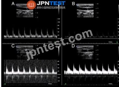
- A. Option B
- B. Option D
- C. Option C
- D. Option A
正解:A
解説:
Option B demonstrates appropriate spectral Doppler gain. Appropriate gain settings ensure that the Doppler signal is adequately amplified without introducing excessive noise or artifacts. In Option B, the spectral waveform is clearly visible with distinct borders, and the background noise is minimal. In contrast, other options might show either under-gained (too little signal) or over-gained (excessive noise and signal) images, making it difficult to accurately interpret the spectral Doppler information. Reference:
ARDMS Sonography Principles and Instrumentation guidelines
"Understanding Ultrasound Physics" by Sidney K. Edelman
質問 # 74
Which change was made after acquiring image A to produce image B?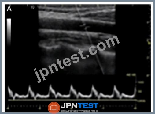
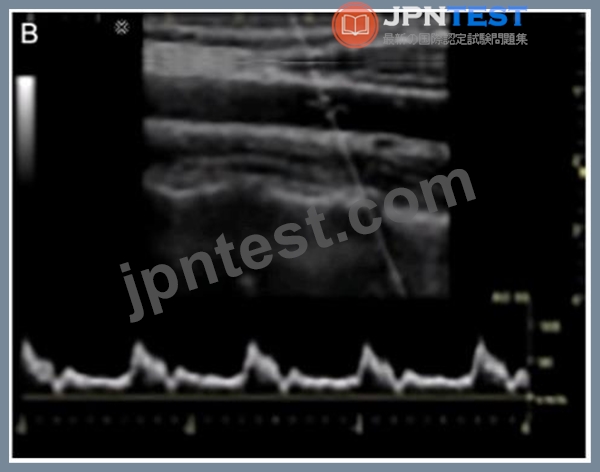
- A. Increased spectral gain
- B. Increased sweep speed
- C. Decreased pulse repetition frequency
- D. Decreased wall filter
正解:C
解説:
Increased Sweep Speed: This affects the display of the waveform over time but does not impact the appearance of the spectral Doppler signal in the way shown.
Decreased Pulse Repetition Frequency (PRF): Lowering the PRF can lead to aliasing, which is evident as the waveform wrapping around in the spectral display from image A to image B. This makes the velocity appear higher than it actually is.
Decreased Wall Filter: This adjustment primarily affects the elimination of low-frequency Doppler signals but does not typically cause the kind of changes seen in the images.
Increased Spectral Gain: Increasing the gain would result in a brighter spectral display but not the wrapping of the signal as seen.
Reference:
"Understanding Ultrasound Physics" by Sidney K. Edelman
ARDMS Sonography Principles and Instrumentation study materials
質問 # 75
At which angle to blood flow would the maximum Doppler shift occur?
- A. 0 degrees
- B. 90 degrees
- C. 30 degrees
- D. 60 degrees
正解:A
解説:
The Doppler shift is highest when the angle between the ultrasound beam and the direction of blood flow is 0 degrees. This is because the cosine of 0 degrees is 1, maximizing the Doppler frequency shift. As the angle increases towards 90 degrees, the cosine value decreases, reducing the Doppler shift.
Reference:
ARDMS Sonography Principles and Instrumentation guidelines
Hoskins, P. R., Thrush, A., Martin, K., & Whittingham, T. A. (2010). Diagnostic Ultrasound: Physics and Equipment.
質問 # 76
......
全問SPI問題集とSonography Principles and Instrumentationトレーニングコース:https://www.jpntest.com/shiken/SPI-mondaishu
無料テストエンジンSonography Principles and Instrumentation認定試験:https://drive.google.com/open?id=1KbSa8CIh5bgwCwzYvrbRM3fuP0Qjgizy